
Tooth Anatomy Poster on Behance
A chart is a diagrammatic representation of the teeth showing all the surfaces of the teeth. The charts in the examination will be used to show: Teeth present Teeth missing Work to be carried out Work completed Surfaces with cavities and restorations etc. When charting, the mouth is looked on as being a flat line.

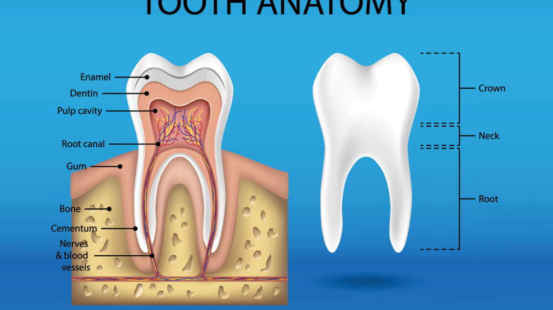
Structure of Tooth Diagram
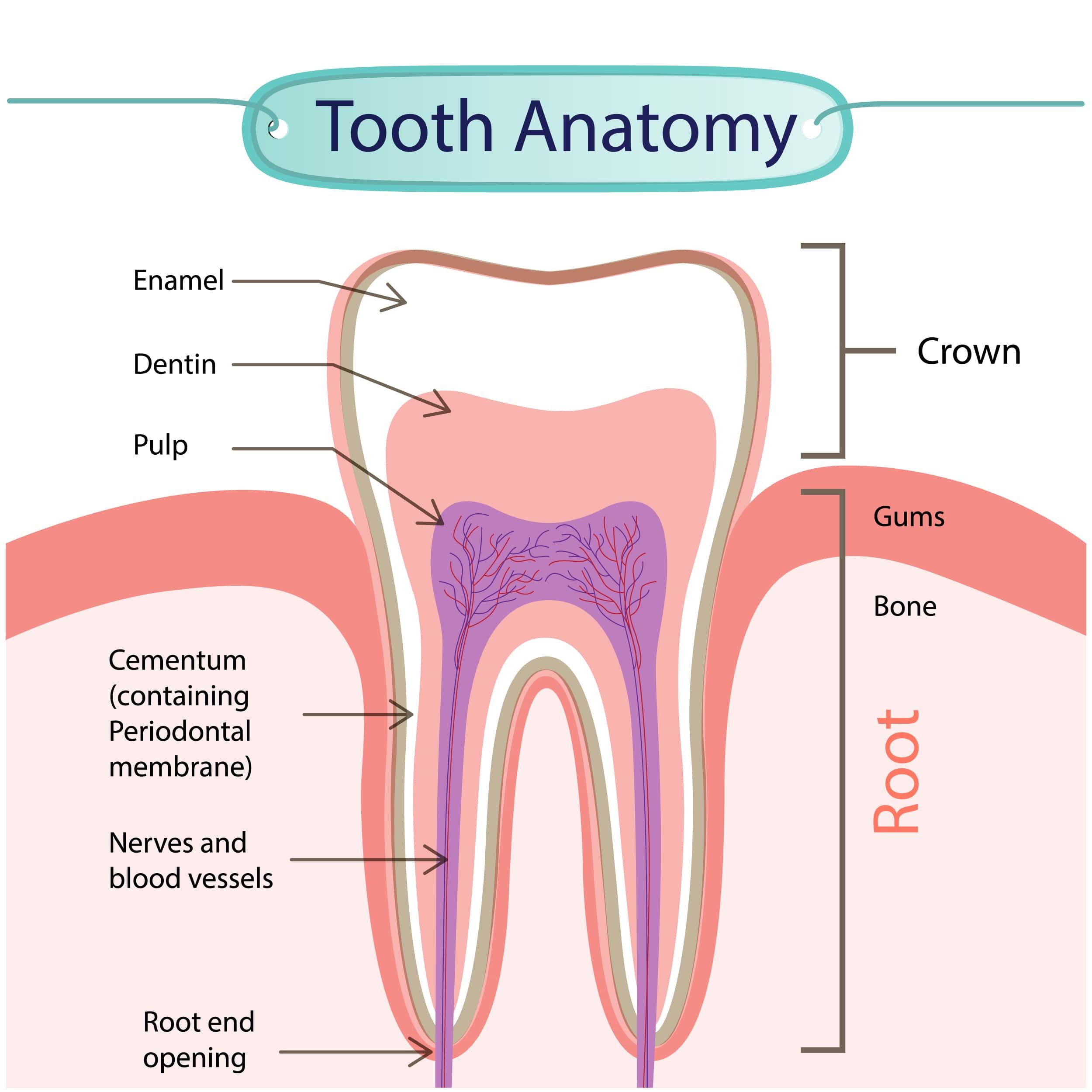
Identifying the teeth. The numbering system shown is the one most commonly used in the US. Each tooth has a crown and a root. The canines have the largest and strongest roots. An inner pulp contains blood vessels, lymphatics, and nerves, surrounded by the hard but porous dentin, which is sensitive to touch and to temperature changes.
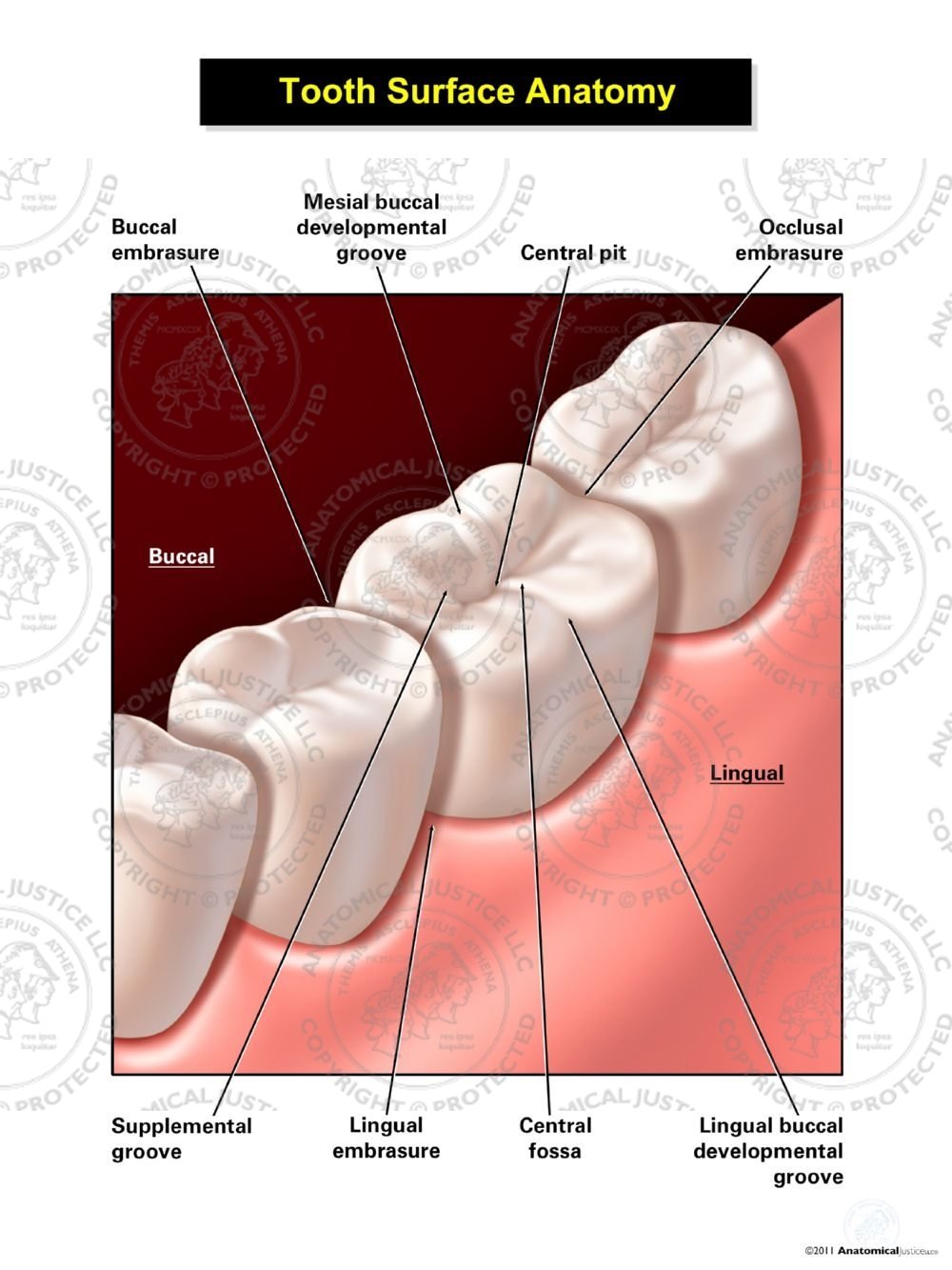
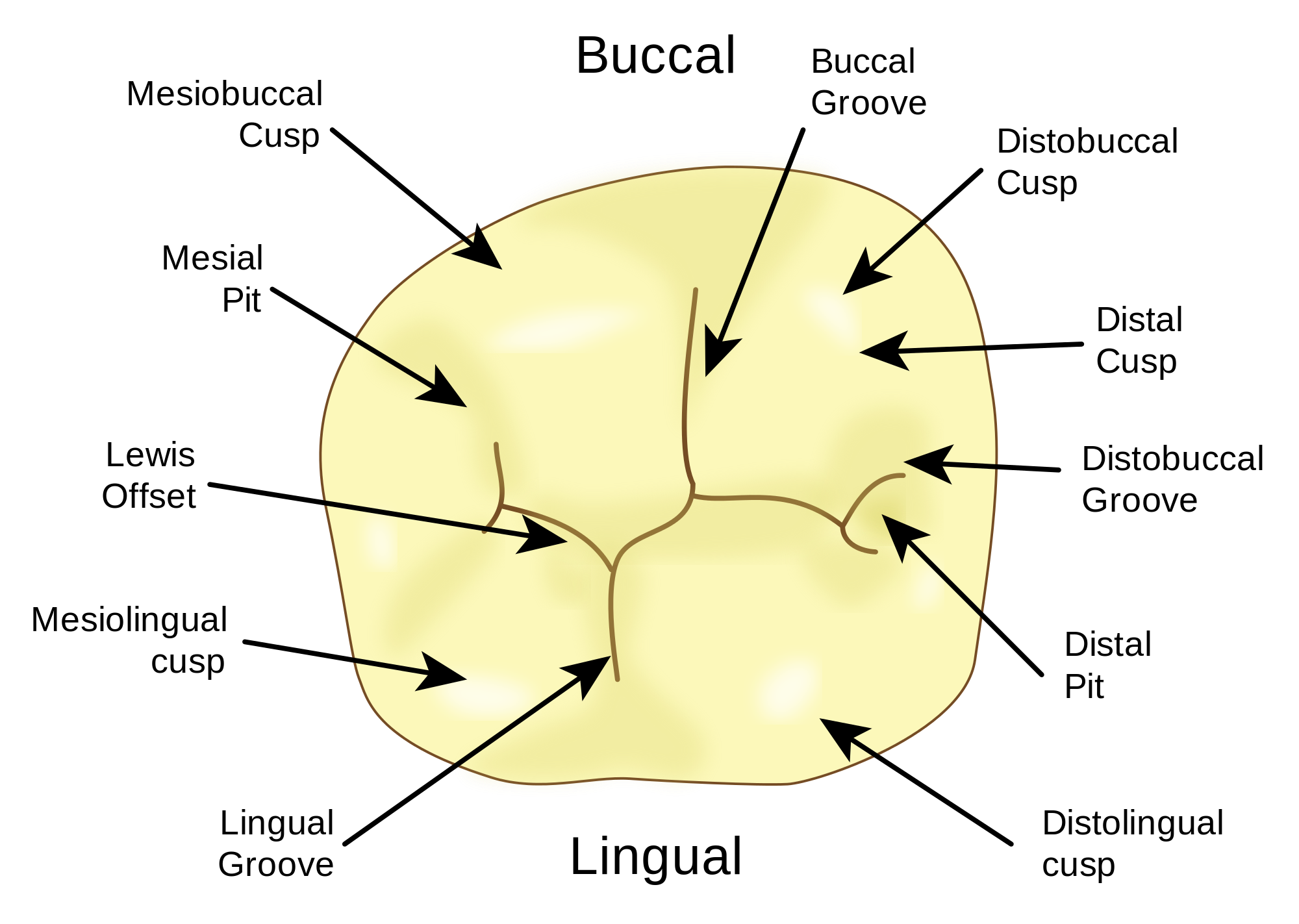
Tooth Surface Anatomy
Interproximal portions of the teeth were the most represented diseased surfaces, while tooth mid surfaces (buccal or lingual) were the less represented ( Figure 1 and Table 1).

Human Teeth Structure With Labels Ilustración de stock Getty Images
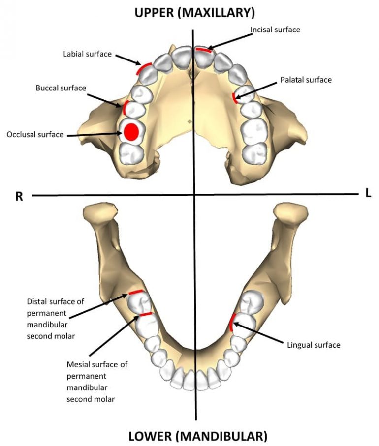
Structure of the tooth Articulating surfaces Crown Root Clinical aspects Sources + Show all Types of teeth The teeth are divided into four quadrants within the mouth, with the division occurring between the upper and lower jaws horizontally and down the midline of the face vertically.
Your Tooth Surfaces Explained Dental Clinique
A teeth chart is a simple drawing or illustration of your teeth with names, numbers, and types of teeth. There are separate teeth number charts for adults as well as babies. This diagram helps us learn the names of each tooth, the corresponding number, and their location.

Very colorful, userfriendly poster covering the Anatomy of the Teeth this large central image
Dental anatomy is a field of anatomy dedicated to the study of human tooth structures. The development, appearance, and classification of teeth fall within its purview. (The function of teeth as they contact one another falls elsewhere, under dental occlusion.)Tooth formation begins before birth, and the teeth's eventual morphology is dictated during this time.

Introduction to Dental Anatomy (Dental Anatomy, Physiology and Occlusion) Part 2
deciduous teeth - The first set of teeth, also known as primary teeth. dentin - The tissue of the tooth between the pulp and the enamel and cementum; the majority of the tooth. dentition - A set of teeth. dentoenamel junction (DEJ) - The junction where the enamel meets the dentin. distal - Surface of the tooth away from the midline of.

Printable Tooth Surface Chart Customize and Print
Your teeth are divided into four types: incisors, cuspids, premolars and molars. Although most people have 32 permanent teeth, Health Direct says that we start with just 20 baby teeth, which include only incisors, canines and molars. Getting a little brush up—pun intended—on all the human teeth names, each type's location and their function can help you better understand why your oral care.
Tooth Anatomy Infographic Smile Angels of Beverly Hills
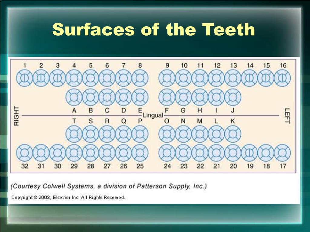
A. Right maxillary 2nd primary molar B. Right maxillary 1st primary molar C. Right maxillary cuspid D. Right maxillary lateral E. Right maxillary central T. Right mandibular 2nd primary molar S. Right mandibular 1st primary molar R. Right mandibular cuspid Q. Right mandibular lateral P. Right mandibular central F. Left maxillary central

The Different Types of Teeth Mortenson Family Dental
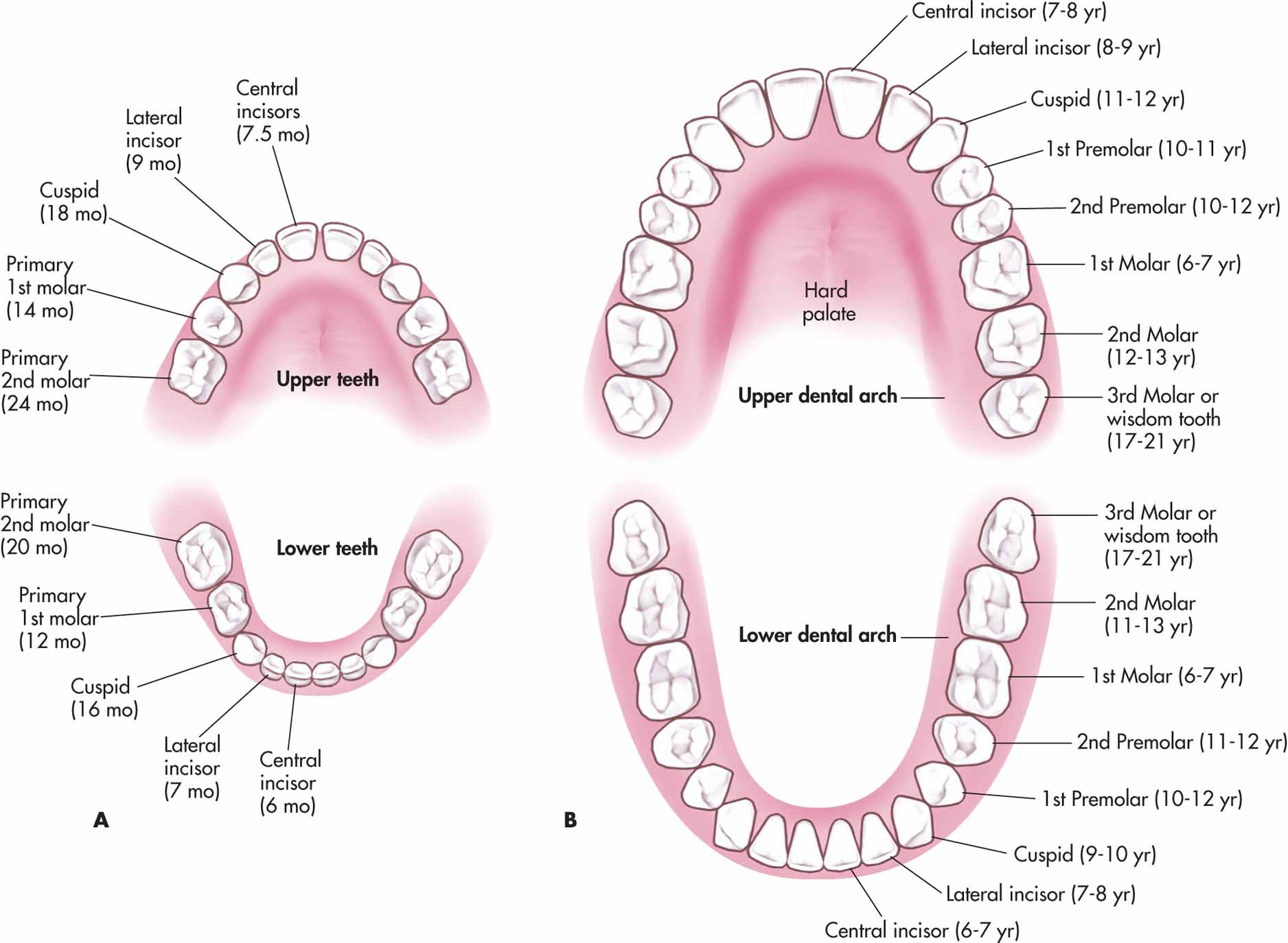
Teeth names and numbering There are thirty-two teeth in total in the oral cavity of an adult dentition. One half, or sixteen, are embedded in the maxilla, while the lower half are situated within the mandible.The name of teeth on each arcade is self-explanatory - the top sixteen are named 'maxillary teeth', while the bottom half are named 'mandibular teeth'.

Printable Tooth Surface Chart Customize and Print
What is dental charting? Dental charting is a process in which your dental healthcare professional lists and describes the health of your teeth and gums. Periodontal charting, which is a part of.

Illustration showing the maxillary and mandibular dental arches. The... Download Scientific
Incisal - The biting edge of an anterior tooth. Lingual - The surface that faces the tongue. Mesial - The surface that is closest to the midline of the face. Occlusal - The chewing surface of posterior teeth. Proximal - Tooth surfaces that are next to each other (i.e., distal of lateral incisor and mesial of canine). Figure 6.
Teeth Types of Teeth, Tooth Anatomy Clinical Relevance Geeky Medics
In the human mouth, teeth make up roughly 20% of the total surface area of the oral cavity. There are 2 types of dentition that develop in humans: Primary (colloquially termed baby or milk) teeth of which there are 20 in total, made up of 8 incisors, 4 canines and 8 molars.
The Anatomy of Your Teeth
The Tooth Chart. This area displays a graphical representation of the teeth in the patient's upper and lower jaws, including Base Charting, Historical Charting and Planned Treatment. The Tooth Chart can be displayed in two modes: Two Dimensional: Three Dimensional: 3D charting is explained in detail later in this document.
Deciduous And Permanent Teeth and Structure of a Tooth Earth's Lab
Dental treatment documentation and billing require to properly identify teeth and tooth surfaces. Incorrect TID and SID are frequent reasons for claim denial and charting errors. We use the universal numbering system for tooth identification. Here is a link to Wikipedia to explain this system.
PPT Hard Tissue Charting PowerPoint Presentation ID355930
Health tips Types of teeth Most people start off adulthood with 32 teeth, not including the wisdom teeth. There are four types of teeth, and each plays an important role in how you eat,.